Your pet’s body is composed of an intricate network of systems that function together in a delicate balance. Disease upsets this balance, often causing varied and baffling clinical signs that make identifying the primary cause challenging. Our hospital has several board-certified veterinary specialists who focus on diagnosing, treating, and managing complicated medical conditions, such as metabolic diseases or cancer. Reaching a diagnosis often requires an extensive combination of diagnostic tests that allow the internists to start pulling the pieces together to determine a treatment plan. Many diagnostics require advanced equipment and expertise that can only be found at a specialty hospital, such as CASE. Diagnostic tests used by our doctors to reach a diagnosis in complex medical cases may include blood work, ultrasound, diagnostic imaging, and endoscopy.
Blood Work for Pets
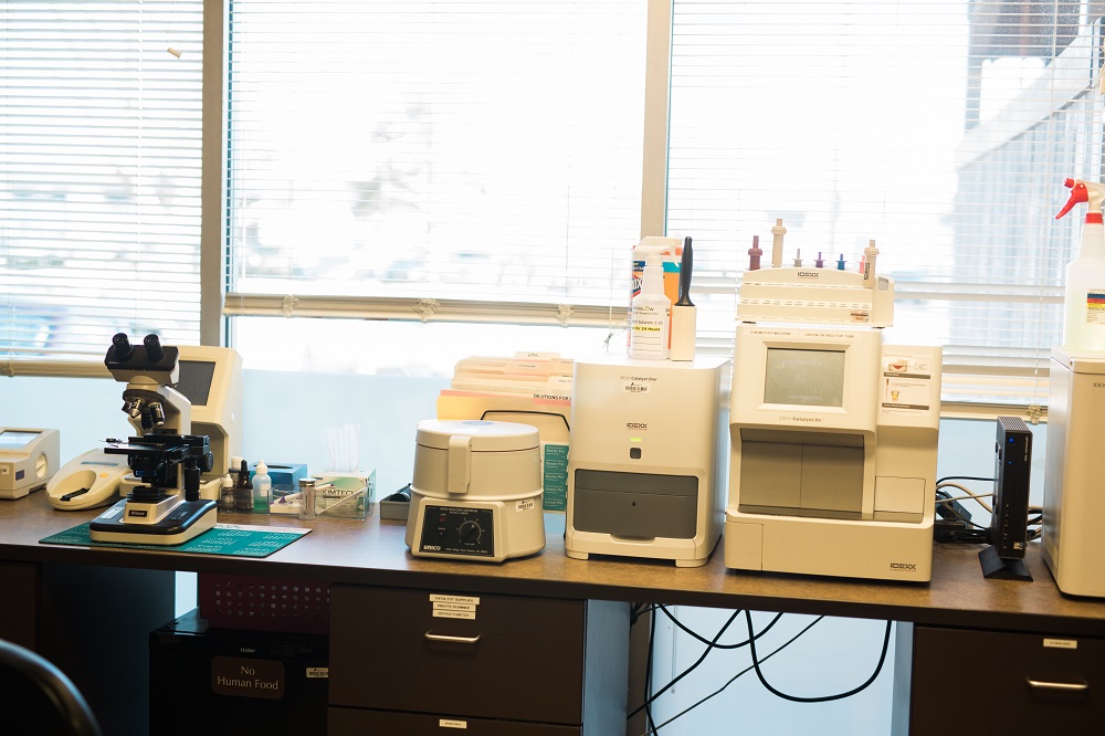
Our diagnostic investigation typically starts with blood testing that provides information about your pet’s overall health status, and often includes:
- Complete blood count (CBC) — A CBC measures the different cellular components of your pet’s blood, including red blood cells, white blood cells, and platelets. Each cell type performs specific functions, and an increase or decrease can indicate a particular problem, such as infection, anemia, or a clotting disorder. All your pet’s blood cells are made by her bone marrow, so abnormal counts can also indicate a bone marrow disorder.
- Blood chemistry — A blood chemistry measures chemicals produced by your pet’s various organs to provide information about their function. Blood chemistry abnormalities can often pinpoint a problem to a specific organ or body system, but further diagnostic testing is often necessary to obtain a more detailed diagnosis.
- Specialized blood tests — Specific blood tests are performed to evaluate individual organs not included in a typical blood chemistry panel. For example, specialized blood tests can assess pancreatic function to aid in a pancreatitis diagnosis.
Blood work often helps us narrow down the body system affected by disease, and then we can use more specialized tests to evaluate specific organs.
Ultrasound for Pets
Ultrasound is a useful diagnostic tool that allows us to view internal body structures in real time. As the ultrasound transducer is moved over your pet’s body surface, sound waves that bounce off internal structures are produced, received, and converted into an image. Ultrasound is neither invasive nor painful, and can often be performed on pets who are awake or only lightly sedated. It’s veterinary medical uses include:
- Identifying abnormal tissue growth, such as cancer
- Guiding needles to collect tissue samples or urine
- Observing heart function (i.e., echocardiogram)
- Identifying urinary stones
- Evaluating gallbladder disease
- Assessing kidney structure
Diagnostic Imaging for Pets
Our diagnostic imaging department offers several additional modalities that allow us to directly view your pet’s internal body structures without using surgical procedures. Imaging techniques that often help us diagnose disease include:
- Digital radiography — Digital radiography allows us to take X-rays of your pet that are transmitted to a computer screen instead of printed on film. X-rays provide excellent bony detail, but minimal soft-tissue detail; however, contrast agents can sometimes be used to provide more information about soft-tissue structures. For example, an oral contrast agent can be administered to pets with gastrointestinal (GI) motility disorders, and serial X-rays taken to evaluate GI function.
- Computed tomography (CT) — A CT uses X-rays to generate thin, cross-sectional slices of a patient’s body that provide better detail than traditional X-rays. CT has a variety of uses, including:
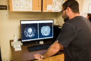 Diagnosis, location, and measurement of cancerous tumors
- Spinal disease diagnosis, such as intervertebral disc disease
- Skeletal injury diagnosis, since tiny bones and surrounding soft-tissue structures, such as blood vessels and nerves, are shown clearly
- Bone and joint disease diagnosis, such as hip or elbow dysplasia
- Portosystemic shunt diagnosis
- Surgical planning, particularly for tumor biopsy or removal
Pets are anesthetized for a CT scan, because they must remain completely still throughout the procedure, and are closely monitored by our trained technicians while under anesthesia. Most CT scans take less than 30 minutes, so your pet will be awake and back on her feet in no time.
Endoscopy for Pets
An endoscope, which is a camera mounted on the end of a rigid or flexible tube, can be inserted into your pet’s natural body openings, such as her mouth or nose, or through an incision made into her thorax, abdomen, or a joint. The high-definition camera allows our internists to view internal body structures up close, in color, and in real time. Our surgery and internal medicine departments perform a variety of endoscopic procedures, including:
- GI endoscopy — An endoscope can be inserted through your pet’s mouth, and advanced through her esophagus and into her stomach to view internal structures, collect tissue samples, and remove foreign bodies.
- Arthroscopy — Arthroscopy is the endoscopic examination of joint surfaces. An incision is made into a pet’s joint, and the endoscope is introduced to visualize joint surfaces in greater detail than other imaging modalities can provide.
- Bronchoscopy — Pets with chronic coughing or difficulty breathing may require endoscopic examination of their airways. During the procedure, a bronchoalveolar lavage (BAL) is often performed to collect a sample from the airway surface for microscopic evaluation and bacterial culture to identify the cause of chronic airway disease.
- Rhinoscopy — An endoscope can be inserted into your pet’s nose to diagnose the cause of chronic nasal discharge. Nasal polyps and foreign bodies, two common culprits, can often be removed with a grasping tool introduced through the endoscope tubing.
Once individual test results are integrated to reach a diagnosis, our specialists will design a treatment plan for your pet. The same tools used to diagnose her disease are often used to monitor treatment response and disease management.
Contact us if your family veterinarian has referred your pet to one of our doctors for diagnosis of a challenging medical condition.