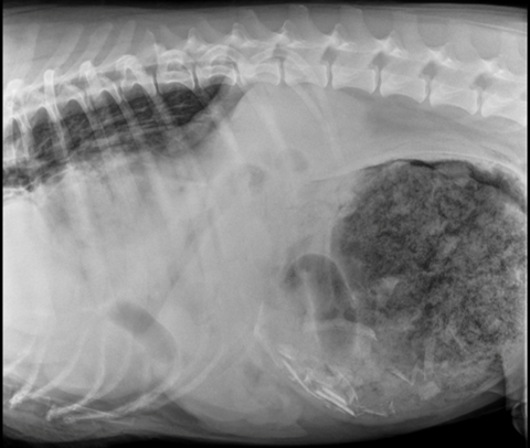
Bunsen, a 3-year-old Bernese Mountain Dog, presented to CASE ER for a distended abdomen and appearing extremely uncomfortable. His heart rate was elevated and his abdomen was extremely firm on palpation. Radiographs of Bunsen’s chest and abdomen showed two issues at hand. Bunsen had developed gastric dilatation and volvulous (GDV), but also had a diaphragmatic hernia (a communication between the chest cavity and abdomen due to a rent in the diaphragm, resulting in abdominal contents that migrated into the chest cavity).
Bunsen was stabilized and prepped for surgery by the CASE Emergency Service and proceeded to emergency surgery with Dr. Kyle Martin. In surgery, Bunsen’s stomach was de-rotated and the abdominal contents within the chest cavity were retrieved. One section of bowel in the chest cavity had poor color and blood flow, thus requiring resection and suturing the remaining healthybowel back together. Lastly, the foreign material within the stomach was removed and Bunsenreceived a gastropexy (tacking the stomach to the abdominal wall) to prevent another GDV from occurring in the future.
Upon closing the diaphragm and abdominal cavity, the chest cavity continued to produce air at a constant rate, thus a chest tube was placed to help evacuate the air and fluid building up following removal of the abdominal contents. Bunsen required blood pressure supportive medications during surgery along with mechanical ventilation. He recovered from anesthesia and was able to be extubated.
Bunsen’s care was continued with the CASE Critical Care Team. He continued to have a large amount of air accumulating within his chest cavity called a continuous pneumothorax. A second chest tube was placed to help evacuate the air and he was placed on continuous suction to facilitate negative pressure within the chest cavity.
A CT scan of the chest cavity was performed and did not reveal an obvious bulla (air pockets within the lung). Bunsen had a blood patch pleurodesis performed, where Bunsen’s own blood was removed and inserted into the chest cavity to help form a fibrous clot (“patch”) and prevent further leaking from the lungs. It was suspected that the abdominal contents within the chest cavity developed adhesions to the lungs, and when the abdominal contents were removed, there was likely trauma to the lungs creating the continuous air accumulation within the chest cavity. The goal with the blood patch pleurodeses was to hopefully prevent Bunsen from going back to surgery to explore the chest cavity.
Following the first blood pleurodesis, air was still accumulating within the chest cavity, albeit at a slightly slower rate. A second blood patch pleurodesis was performed 24 hours later with the same goal of preventing surgery to explore the chest cavity. Success was achieved with the second blood patch and his chest cavity was no longer producing continuous air. In conjunction, Bunsen also developed abdominal pain in the post-operative period, which was attributed to pancreatitis from trauma to the pancreas sustained from the GDV and corrective surgery. Bunsen received local regional anesthesia by the CASE Critical Care Team which did improve his overall comfort.
Unfortunately, with the systemic inflammation present due to multiple factors (surgery and pancreatitis), Bunsen developed a large clot (thrombus) within his heart. This is not an uncommon phenomenon given the degree of inflammation present within the body. Where there is inflammation, there will be an increased propensity for the body to form clots. Bunsen was started on anticoagulants immediately upon discovery of the thrombus. The clot was present within the right atrium of the heart and eventually dislodged into the main pulmonary artery. The risk of dislodgement of a thrombus is a massive pulmonary thromboembolism that can have detrimental effects on the function of the heart and prevents blood from reaching the lungs to become oxygenated. Fortunately for Bunsen, the thrombus did not develop a complete occlusion of the main pulmonary artery and his body was able to compensate for this change.
Bunsen had a temporary esophagostomy tube placed prior to discharge to help facilitate nutritional support, medication administration and maintenance of hydration. He continued to make improvements and was discharged after 7 days in hospital. Bunsen has made a remarkable recovery over the past month and highlights the integrative team at CASE. Bunsen is always happy to come visit the team at CASE.